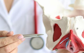
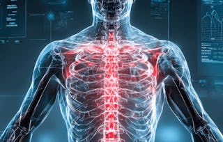
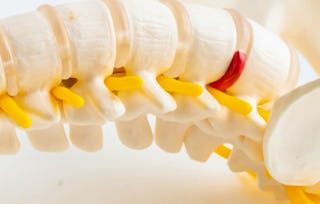
This course teaches learners the underlying principles behind conventional radiography, computerized axial tomography (CT), magnetic resonance imaging (MRI), and ultrasound. The radiology of chest, abdomen, pelvis, extremities, spine and brain are taught in this course using a combination of lectures and extensive practical activities and assessments.
Visualizing the Living Body: Diagnostic Imaging
Visualizing the Living Body: Diagnostic Imaging


Instructors: William B. Stewart
Access provided by Universidad Austral
43,344 already enrolled
Gain insight into a topic and learn the fundamentals.
388 reviews
Intermediate level
Recommended experience
Flexible schedule
1 week at 10 hours a week
Learn at your own pace
93%
Most learners liked this course
Details to know

Shareable certificate
Add to your LinkedIn profile
Assessments
7 assignments
Taught in English
See how employees at top companies are mastering in-demand skills

There are 7 modules in this course
Instructors
Instructor ratings
(133 ratings)Offered by
Why people choose Coursera for their career

Felipe M.
Learner since 2018
"To be able to take courses at my own pace and rhythm has been an amazing experience. I can learn whenever it fits my schedule and mood."

Jennifer J.
Learner since 2020
"I directly applied the concepts and skills I learned from my courses to an exciting new project at work."

Larry W.
Learner since 2021
"When I need courses on topics that my university doesn't offer, Coursera is one of the best places to go."

Chaitanya A.
"Learning isn't just about being better at your job: it's so much more than that. Coursera allows me to learn without limits."
Learner reviews
- 5 stars
83.76%
- 4 stars
14.69%
- 3 stars
1.28%
- 2 stars
0%
- 1 star
0.25%
Showing 3 of 388
HA
Reviewed on Aug 1, 2024
Amazingly delivered concepts !!!! Impressed by all the professors of Yale University
LJ
Reviewed on Apr 7, 2024
This course was thorough in teaching the basic interpretation of normal radiology images, I have learnt a lot and excited to apply these skills in my field.
MR
Reviewed on Apr 24, 2024
"Excellent! The optimal pace and clarity of the content significantly enhanced the learning experience, making it exceptionally efficient."
Explore more from Health
Yale University
University of Glasgow
Korea Advanced Institute of Science and Technology(KAIST)
Yale University